Retinal Imaging
Retinal Imaging
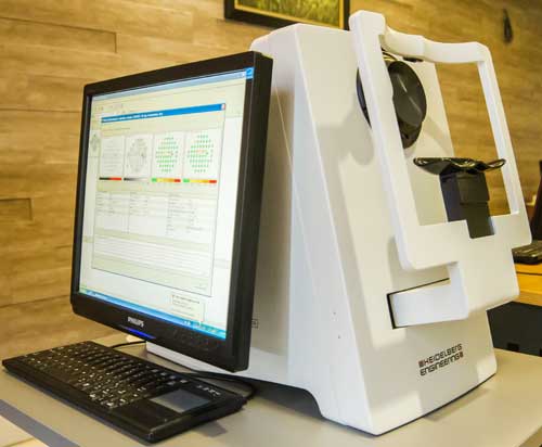
HEP
Visual field testing remains the main diagnostic tool to provide information about a patient’s visual function, quality of vision, and quality of life. Functional testing supports clinicians in early disease detection, staging of disease, or trend analysis in progressive glaucoma. 2-in-1 perimetry with HEP offers the most appropriate test for each stage of the disease. It allows you to offer the right stimulus to your patient at the appropriate stage of the disease.(www.heidelbergengineering.com)
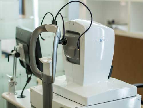
Optical Coherence Tomography (OCT)
Optical coherence tomography (OCT) is a non-invasive imaging test that uses light waves to obtain detailed images from the retina and to evaluate disorders of the optic nerve. It helps ophthalmologists in the diagnosis of glaucoma and retinal diseases like age-related macular degeneration and diabetic retinopathy.
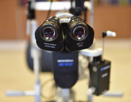
Slit Lamp Photography
It is an instrument consisting of a high-intensity light source and a special microscope that facilitates an examination of the anterior segment and posterior segment of the human eye including the eyelid, the sclera, the conjunctiva, the iris, the natural crystalline lens, and the cornea. Slit-lamp is usually used during a regular checkup to examine your eyes for any diseases or abnormalities. It also has a second lens that is used to evaluate the retina.
Ocular Ultrasonography
It is a safe, non-invasive, and easy-to-use diagnostic tool for visualization of ocular pathology and anatomy using high-frequency sound waves. The sound waves travel through the eye and their reflections provide a picture of the structure of the eye. The test has two types namely A-scan and B-scan. A-scan determines the power of the implanted lens before cataract surgery and B-scan is used for looking at spaces behind the eye. Ultrasonography is also used to evaluate the iris, the lens, the ciliary body, and the orbital structures. it is most useful in the condition of opaque ocular media.