Corneal Imaging
Corneal Imaging
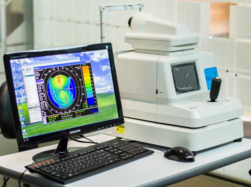
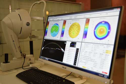
Topography
Topography provides both a qualitative and quantitative evaluation of corneal curvature. It does so by utilizing concentric rings, which project onto the cornea to create a virtual image. The device compares this image to the target size, and the computer then calculates the corneal curvature. Although many different systems are available, all share some unifying measurement characteristics. The computerized topographer can generate various graphical representations when performing corneal mapping for the diagnosis of conditions and/or contact lens fitting.
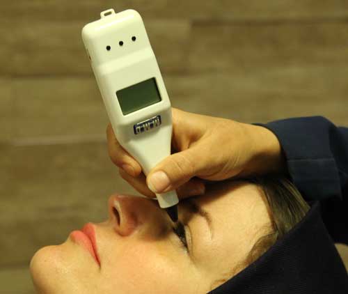
Pachymetry
Pachymetry is a measurement of the thickness of the cornea. Some conditions can cause the cornea to thicken or fill with liquid. Pachymetry can be a vital tool in diagnosing such conditions in order to limit the loss of vision.
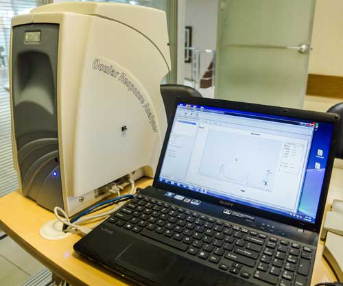
ORA
The ocular response analyzer (ORA) is a non-contact (air puff) tonometer that does not require topical anesthesia and provides additional information on the biomechanical properties of the cornea. It uses an air pulse to deform the cornea into a slight concavity. The difference between the pressures at which the cornea flattens inward and outward is measured by the machine and termed corneal hysteresis (CH). The machine uses this value to correct the effects of the cornea on measurement.
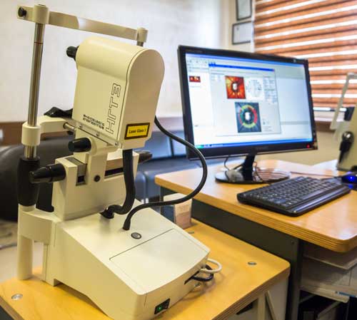
HRT-Cornea-Module
- Early clinical assessment of Keratitis
•Clinical assessment of the nerve plexus with diabetic neuropathy
•Gauge keratoplasty & DSAEK immune response
•Identify distinct dystrophy subtypes
•Endothelial cell counting
The Heidelberg Retina Tomograph (HRT) is a proven, essential tool for detecting and managing glaucoma, especially for assisting in the identification of pre-perimetric disease and tracking progression. The Ancillary Study to the Ocular Hypertension Treatment Study (OHTS) demonstrated that optic disc analysis detected glaucoma conversion in 55% of cases, before any detectable loss of visual function. The HRT has proved to be a robust predictor of glaucoma. OHTS also concluded that the HRT recognized temporal superior defects which later developed into confirmed glaucoma in 40% of cases – looking only at baseline measurements – then confirmed the diagnosis with an average of 5 years of follow-up. Equally impressive was that 93% of all HRT cases flagged as within normal limits at baseline remained within normal limits during the same time period. (www.heidelbergengineering.com)
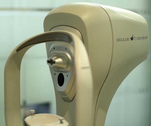
Corvis
The Corvis® ST is a Non-Contact-Tonometer and allows seeing the reaction of the cornea on an air impulse. A high-speed Scheimpflug-camera (4330 frames/sec) records the movements of the cornea which then are displayed on the built-in control panel in ultra-slow-mo. So far unseen pictures allow exciting new ways in the assessment of corneal properties for various applications from Keratoconus screening to Glaucoma. ( www.oculus.de)
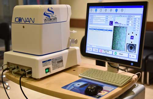
Specular Microscopy
A noninvasive photographic technique that allows you to visualize and analyze the corneal endothelium. Using computer-assisted morphometry, modern specular microscopes analyze the size, shape, and population of endothelial cells. The instrument projects light onto the cornea and captures the image that is reflected from the optical interface between the corneal endothelium and the aqueous humor. The reflected image is analyzed by the instrument and displayed as a specular photomicrograph. In clinical practice, specular microscopy is the most accurate way to examine the corneal endothelium.
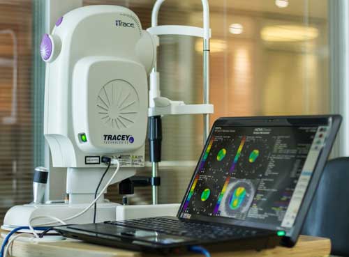
ITrace
ITrace offers integrated corneal topography and wavefront aberrometry for unique analysis separating corneal and internal aberrations of the eye – a complete function analyzer that can improve surgical planning and help determine the best vision correction treatment for your patients.
•Select the correct aspheric IOL
•Improve treatment outcomes for refractive, AK’s, accommodative & multifocal IOL’s,
•Analyze refractions on a multi-zone basis
•Assess highly aberrated eyes in the most complex cases
•Track visual function over time
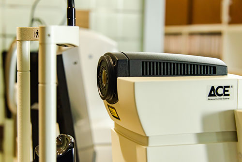
ACE (Advanced Corneal Explorer)
ACE combines the swept-source OCT imaging technology with corneal imaging to enhance diagnostic confidence and accuracy for cataract surgery, refractive surgery, and follow-up of corneal pathologies. ACE is intended for visualization and measurements of the anterior segment of the eye and the measurement of axial length and analysis of the eye.
Pentacam
The Pentacam measurement process takes less than two seconds and minute eye movements are captured and corrected simultaneously. By measuring true elevation points, precise representation, repeatability, and analysis are guaranteed. These data points are then used to generate corneal maps used for diagnosis and treatment.
Pentacam AXL Wave
It is the first device that combines Scheimpflug tomography with optical biometry, total wavefront, objective refraction, and retroillumination. It provides reliable data that are necessary for pre-op assessment and post-op evaluation of cataract and refractive surgeries.
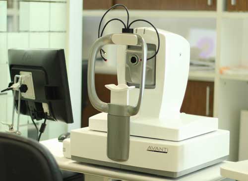
Optical Coherence Tomography (OCT)
Optical coherence tomography (OCT) is a non-invasive imaging test that uses light waves to obtain detailed images from the retina and to evaluate disorders of the optic nerve. It helps ophthalmologists in the diagnosis of glaucoma and retinal diseases like age-related macular degeneration and diabetic retinopathy.
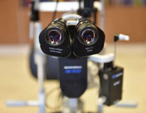
Slit Lamp Photography
It is an instrument consisting of a high-intensity light source and a special microscope that facilitates an examination of the anterior segment and posterior segment of the human eye including the eyelid, the sclera, the conjunctiva, the iris, the natural crystalline lens, and the cornea. Slit-lamp is usually used during a regular checkup to examine your eyes for any diseases or abnormalities. It also has a second lens that is used to evaluate the retina.