Other Diagnostic Services
Other Diagnostic Services
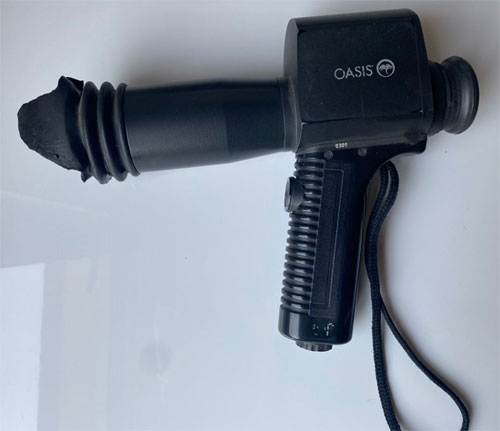
UBM
Ultrasound biomicroscopy utilizes high-frequency transducers to obtain high resolution. Our unit, with a 50 MHz transducer, achieves a resolution of approximately 50 microns and has a tissue penetration of 4-5 mm. Scanning is performed with the patient in the supine position under standardized room lighting conditions. Scanning is performed under topical anesthesia using a 20 mm eye cup which is inserted between the lids and filled with saline solution. In vivo, cross-sectional or transverse images can then be obtained detailing the cornea
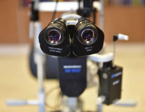
Slit Lamp Photography
It is an instrument consisting of a high-intensity light source and a special microscope that facilitates an examination of the anterior segment and posterior segment of the human eye including the eyelid, the sclera, the conjunctiva, the iris, the natural crystalline lens, and the cornea. Slit-lamp is usually used during a regular checkup to examine your eyes for any diseases or abnormalities. It also has a second lens that is used to evaluate the retina.
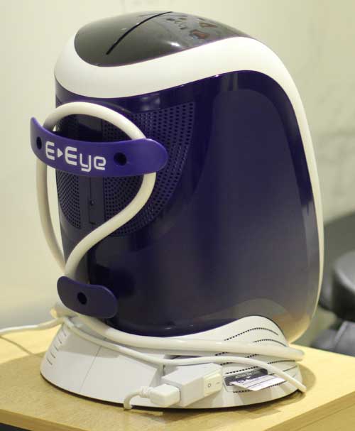
E-Eye
It is a medical device for the treatment of dry eye using intense regulated pulsed light technology. A series of 5 flashes –IRPL is performed under each eye in the areas (suborbital and zygomatic region) where the parasympathetic nerve passes to prevent or treat meibomian gland dysfunction and dry eye syndrome.
Pupilometery
It is a handheld device that provides accurate, reliable, and objective pupil size and reactivity data. It measures the distance between pupils through visual stimuli.
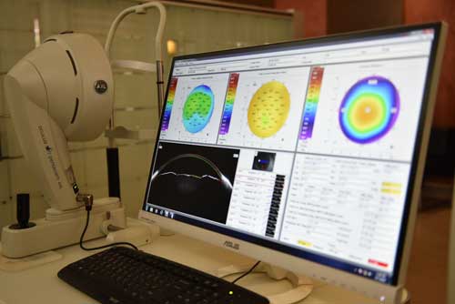
Pentacam
The Pentacam measurement process takes less than two seconds and minute eye movements are captured and corrected simultaneously. By measuring true elevation points, precise representation, repeatability and analysis are guaranteed. These data points are then used to generate corneal maps used for diagnosis and treatment.
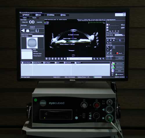
Ocular Ultrasonography
It is a safe, non-invasive, and easy-to-use diagnostic tool for visualization of ocular pathology and anatomy using high-frequency sound waves. The sound waves travel through the eye and their reflections provide a picture of the structure of the eye. The test has two types namely A-scan and B-scan. A-scan determines the power of the implanted lens before cataract surgery and B-scan is used for looking at spaces behind the eye. Ultrasonography is also used to evaluate the iris, the lens, the ciliary body, and the orbital structures. it is most useful in the condition of opaque ocular media.